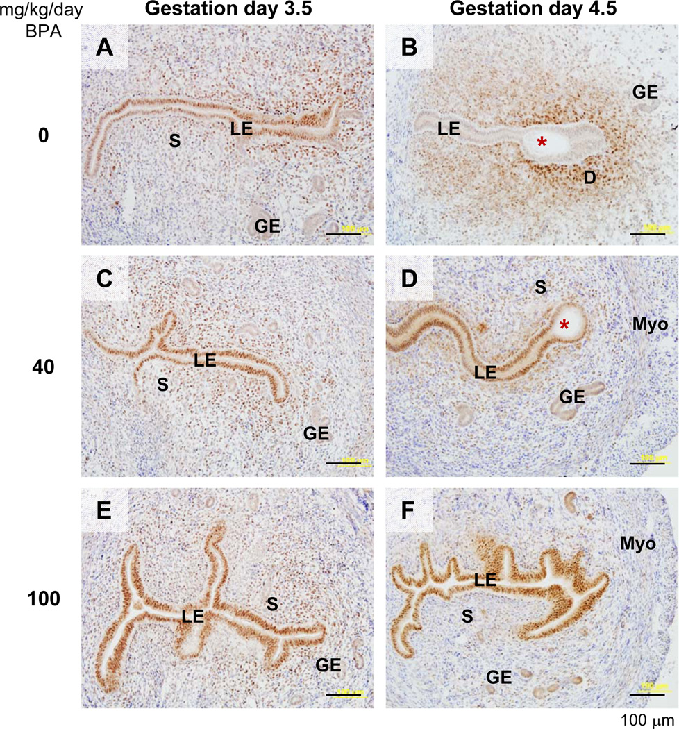
Figure 4.
Immunohistochemical detection of progesterone receptor (PR) in gestation day 3.5 and day 4.5 uteri upon preimplantation BPA treatment. Uterine cross sections (10 µm) were processed for detecting PR localization (brown staining). A representative section was from each of the following groups: A. Gestation day 3.5, 0 mg/kg/day BPA (control). B. Gestation day 4.5, 0 mg/kg/day BPA (control). C. Gestation day 3.5, 40 mg/kg/day BPA. D. Gestation day 4.5, 40 mg/kg/day BPA. E. Gestation day 3.5, 100 mg/kg/day BPA. F. Gestation day 4.5, 100 mg/kg/day BPA. No specific staining was detected in the negative control (data not shown). Red star, embryo; LE, luminal epithelium; S, stroma; GE, glandular epithelium; D, decidual z one; Myo, myometrium. Scale bar: 100 µm.

