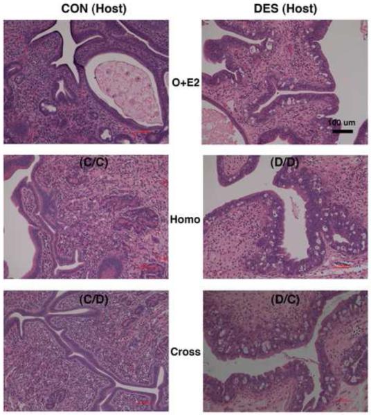
Figure 7. Effects of neonatal DES treatment on uterine histomorphology in mature, ovariectomized, and estrogen-replaced hamsters and in both groups of host hamsters with viable ovarian transplant tissue masses.
Shown are hematoxylin and eosin-stained uterine cross sections (all taken at the same magnification) from mature (2-mo) animals that: 1) on the day of birth (d-0) were not (control or CON, left panels) or were treated with DES (right panels) and then were prepubertally (d-21) ovariectomized and either 2) received an estradiol-releasing implant (O+E2, upper panels) or received cheek pouch transplants of ovaries from 3) the same (Homo, middle panels) or 4) the other (Cross, lower panels) neonatal treatment group of donor animals (see Table 1 for further transplant group descriptions). Scale bar in upper right panel represents 100 μm.

