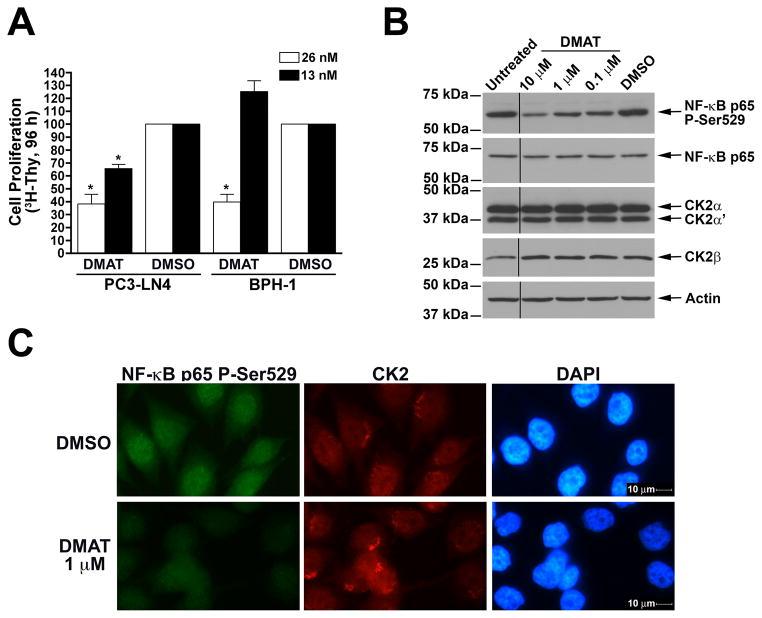
Fig. 1.
Cellular effects of naked DMAT in benign and malignant prostate cancer cells. A. Reduced cellular proliferation following treatment with naked DMAT. PC3-LN4 and BPH-1 cells grown on tenascin-C/fibronectin or laminin protein matrix, respectively, in 96-well plates were treated with 26 and 13 nM DMAT. DMSO was used as the control. [3H]-thymidine was added after 72 h, and counts collected at 96 h following drug addition. Results are expressed relative to the DMSO controls, with treatments and cell lines used indicated below. Error bars indicate SEM (n = 4–6). * indicates significant difference from control (P < 0.05). B. Treatment with DMAT results in loss of phospho-Ser529, but not total, NF-κB p65, and no reduction in CK2 expression. PC3-LN4 cells were treated with various concentrations of DMAT as indicated and processed for immunoblot analysis. Treatments and concentration are shown above, with molecular weight markers and proteins detected indicated on the left and right, respectively. Removal of an intervening lane between untreated and 10 μM DMAT is indicated by black line. C. CK2 inhibition by DMAT results in loss of phospho-Ser529 NF-κB p65. PC3-LN4 cells grown on glass coverslips were treated with DMAT or DMSO and fixed 24 h later for indirect immunofluorescence analysis. The treatments applied are indicated on the left and the protein visualized by immunofluorescence or nuclei by DAPI staining is shown above the panels. Magnification, 400×.

