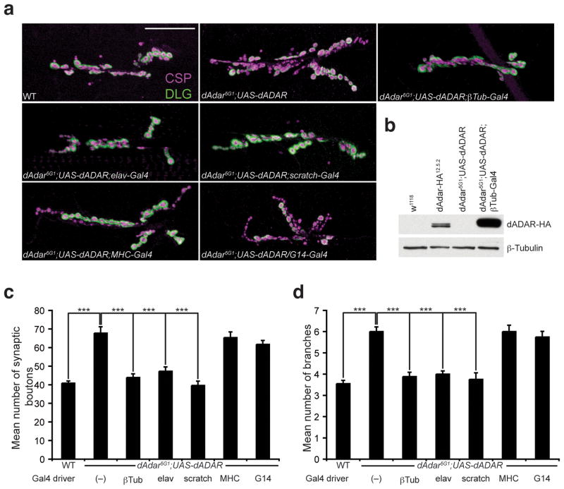
Figure 3. Neuronal expression of dADAR is sufficient for normal NMJ synaptic architecture.
(a) Confocal images of the NMJ from the following genotypes: WT, dAdar5G1 larvae carrying a wild-type UAS-dADAR transgene (dAdar5G1;UAS-dADAR), dAdar5G1;UAS-dADAR;βTub-Gal4 (ubiquitous expression), dAdar5G1;UAS-dADAR;elav-Gal4 (neuronal expression), dAdar5G1;UAS-dADAR;scratch-Gal4 (neuronal expression), dAdar5G1;UAS-dADAR;MHC-Gal4 (muscle expression), and dAdar5G1;UAS-dADAR/G14-Gal4 (muscle expression). Larvae were stained for CSP (presynaptic, magenta) and DLG (postsynaptic, green). No noticeable differences in CSP and DLG intensity were observed across genotypes. Scale bar represents 50 μm. (b) Western analysis on L3 brain extracts from the following genotypes: w1118 (negative control for the HA antibody), dAdar-HA12.5.2 (for endogenous levels), dAdar5G1;UAS-dADAR and dAdar5G1;UAS-dADAR;βTub-Gal4. An antibody against HA was used to detect dADAR-HA expression (upper panel). β-Tubulin (lower panel) was used as a loading control. (c,d) Quantification of the average number of type 1 synaptic boutons (c) and synaptic branching (d) in muscles 6/7 for all genotypes. All images and quantification were performed using abdominal hemisegment A3, muscles 6/7. n≥16 for each genotype. Error bars denote s.e.m. ***p<0.001 analyzed by one-way ANOVA, p<0.0001 overall, Tukey-Kramer post-test. See Supplemental Table 1 for control genotypes.

