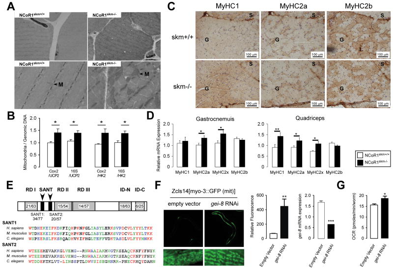
Figure 3. Histological analyses of the muscles of mice and C. elegans.
Transmission electron microscopy of non-oxidative fibers of NCoR1skm+/+ and skm-/- gastrocnemius. M, mitochondria. (B) Relative mitochondrial DNA content (Cox2 or 16S) in gastrocnemius was measured and normalized by genomic DNA content (Ucp2 and Hk) (n=6). (C) Representative MyHC1, 2a, and 2b immunohistochemical detection on serial sections of the soleus and gastrocnemius. (D) Subtypes of MyHCs in gastrocnemius and quadriceps were analyzed by qRT-PCR in NCoR1skm+/+ and skm-/- mice (n=6). (E) Indentity/similarity (%) in the sequences of gei-8 with mammalian NCoR1. Multiple alignment of SANT domains of NCoR1 homologs. Color identifies similarity. (F) Representative pictures of the effects of RNAi-mediated knockdown of gei-8 on mitochondrial morphology and number in a C.elegans strain carrying a mitochondrial GFP-reporter driven by the muscle-specific myo-3 promoter (left panel). Quantification of the mitochondrial induction by fluorescence upon gei-8 knockdown (middle panel), and of the efficacy of the RNAi-mediated gei-8 knockdown by qRT-PCR analysis (right panel). (G) Muscle-specific RNAi inhibition of gei-8 enhances respiration in C.elegans.
Data are expressed as mean ± SEM.
See also Suppl. Fig. 3 and Suppl. Tables 2 and 3.

