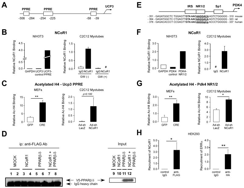
Figure 5. Increased PPARβ/δ and ERR activity in NCoR1skm/- muscle.
(A, B, E, F, and H) NCoR1 recruitment to the PPREs on mouse Ucp3 promoter and to the ERR-RE on human and mouse Pdk4 promoter determined by ChIP in NIH-3T3 cells transfected with an NCoR1-FLAG vector or in C2C12 myotubes. A schematic of the promoters of the Ucp3 (A) and Pdk4 (E) genes and the sequence alignment of the mouse, rat and human Pdk4 promoter is also shown to highlight the conservation of the NR1/2 (or ERR-RE) (E). Boxes indicate putative PPREs in the Ucp3 and the NR1/2, IRS, and Sp1 in the Pdk4 promoter. ChIP experiments for the Ucp3 promoter were performed in C2C12 myotubes both before and 6 hr after addition of a PPARβ/δ agonist (100 nM GW501516). #; not detected. ChIP experiments in HEK293 cells transfected with FLAG-NCoR1 and HA-ERRα vector (H). (C and G) Binding of acetylated histone 4 (H4) to the PPREs on the Ucp3 and to the NR1/2 on the Pdk4 promoters in ChIP assays, using either immortalized NCoR1L2/L2 MEFs, infected with an adenovirus either expressing GFP or Cre recombinase, or C2C12 myotubes infected with the Ad-shNCoR1 virus. Representative data is shown from 3 experiments. (D) Interaction between PPARβ/δ and NCoR1 determined by in vitro co-IP experiments from HEK293 cells, in which NCoR1-FLAG and/or V5-PPARβ/δ are expressed. IP was performed with control IgG (lanes 1, 3, 5, and 7) or anti-FLAG antibody (lanes 2, 4, 6, and 8) and the immunoblot was developed with an anti-V5 antibody. PPARβ/δ co-immunoprecipitated by the anti-FLAG antibody is indicated by an arrow. Input samples are shown in lanes 9 -12.
Data are expressed as mean ± SEM.
See also Suppl. Fig. 6.

