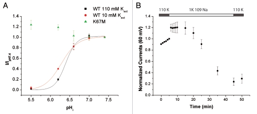
Figure 3.
pH-sensitivity and K+o-regulation in Kir4.1. (A) pHi-sensitivity at different [K+]o. The oocytes were acidified using permeable acetate buffer. pHi was calculated from pHo as described in the methods. The data were fitted with a Hill equation (110 K: pKa = 6.40 ± 0.01, nh = 3.4 ± 0.5, n = 6; 10K: pKa = 6.28 ± 0.01, nh = 2.3 ± 0.4, n = 4; pKa's are significantly different p < 0.0001). (B) Oocytes expressing Kir4.1 K67M were incubated in 110 mM K+ for 1 h prior to start of the TEVC recording. The bath solution was switched to 1 mM K+ at t = 6 min and returned to 110 mM K+ at t = 46 min. Value at t = 50 min: 0.29 ± 0.08 n = 7.

