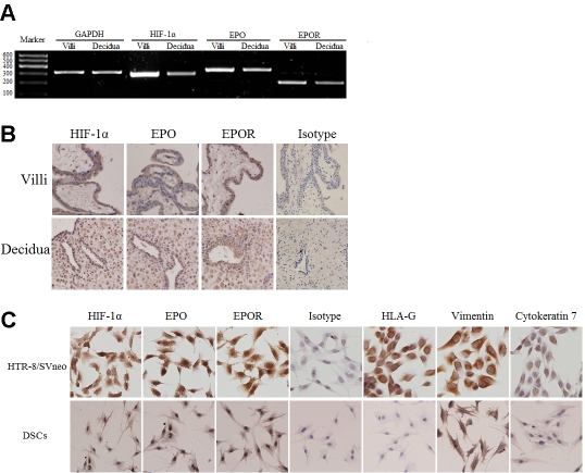
Figure 1.
Expression of HIF-1α, EPO, EPOR at human maternal-fetal interface in the early pregnancy. The expression of HIF-1α, EPO and EPOR on villi and deciduas was detected by RT-PCR (A) and immunohistochemisty (B). The expression of HIF-1α, EPO and EPOR on primary DSCs and HTR-8/SVneo cells was detected by immuocytochemistry (C). HIF-1α, EPO and EPOR were high expressed, and localized to the plasma and nucleus (x200). Trophoblast cells were stained strongly by anti- cytokerat-in7 (CK7) and anti-HLA-G monoclonal antibody (mAb), not by anti-vimentin mAb. Decidual stromal cells (DSCs) were positive for vimentin and negative for CK7 and HLA-G. Magnification: x200. Results were highly reproducible in three independent experiments performed in triplicate.

