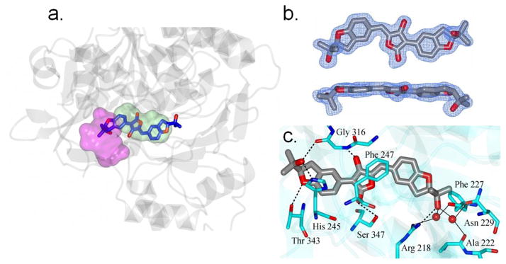
Figure 5. Structure of FLuc containing bound aspulvinone J-CR (5).
a. View of 5 (blue and red cylinders) in the active site of luciferase (grey ribbons). The ATP and luciferin binding regions are colored magenta and green respectively. b. Two views of the Fo-Fc electron density map for 5 contoured at 3sσ. c. Hydrogen bonding between 5 (grey/red) and luciferase (cyan). Water molecules are drawn as red spheres. Direct contacts between luciferase and 5 are shown as dashed lines and water mediated interactions are indicated by the solid lines. Crystallographic data for 5 can be found in Table S2. Coordinates and structure factors have been deposited to the Protein Databank with the accession code 3RIX.

