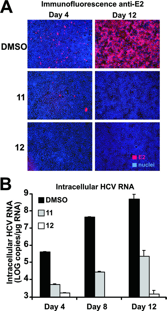
Figure 6. Efficient control of viral spread by selected d,l-α-peptides.
Huh-7 cells were infected at low multiplicity (m.o.i of 0.01) and incubated for the designated periods of time in the presence of 20 µM of the antiviral peptides. Cells were replenished with fresh medium containing the peptides at days 4 and 8 postinfection.
(A) HCV-infected cells are revealed by immunofluorescence microscopy with antibodies against the viral protein E2and are shown in red (E2). Cell nuclei of infected and uninfected cells were stained with Hoescht dye and are shown in blue (nuclei).
(B) Intracellular HCV RNA accumulation was measured by RT-qPCR at the indicated time points. Results are shown as average and mean error of triplicate samples.

