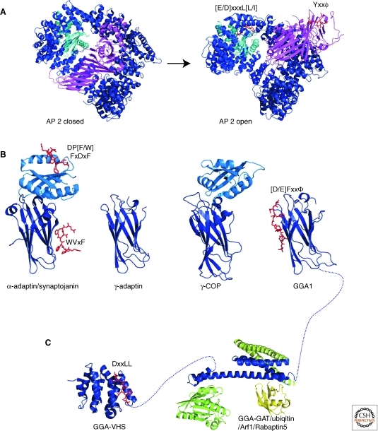
Figure 5.
Structures of adaptor proteins. (A) The AP-2 complex, consisting of α- (light blue), β2- (dark blue), μ2- (magenta), and σ2- (cyan) adaptin, undergoes large-scale conformational changes during its transition from the closed cytosolic form (PDBID 2VGL) to the open, sorting-active form (PDBID 2XA7). (B) The appendage domains of α-adaptin (PDBID 1W80), γ-adaptin (PDBID 1GYU), γ-COP (PDBID 1PZD), and GGA1 (1OM9). (C) The VHS domain of GGA1 (PDBID 1JWG) and the GAT domain (PDBID 1NAF) in complex with Arf1 (light green, PDBID 1J2J), ubiquitin (yellow, PDBID 1WR6), and rabaptin5 (dark green, PDBID 1X79). Bound sorting peptides are shown in red as stick representation and are labeled with the corresponding consensus sequence.

