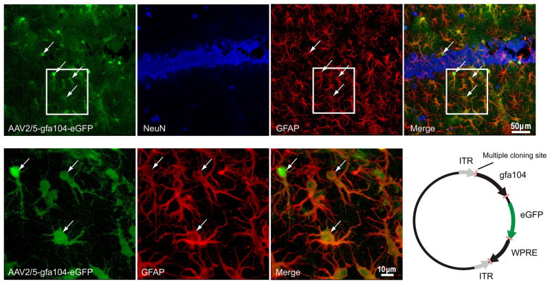
Figure 1. Astrocyte-specific eGFP expression.
(a–d) Confocal images depicting (a) viral injection-induced eGFP expression (green), (b) immunostaining for NeuN (blue), and (c) GFAP (red) in the hippocampus. (d) eGFP co-localizes with GFAP (astrocyte marker; arrows) but not with NeuN (neuronal marker). (e–g) High magnification images show co-labeling of eGFP fluorescence with GFAP staining. (h) Schematic of the AAV2/5-gfa104-eGFP plasmid (see METHODS for details).

