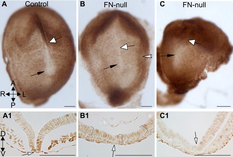
Figure 6. FN is required for the expression of activated forms of SMADs 2/3 at the midline.
A. Control and FN-null embryos (B, C) stained to detect activated forms of SMADs 2 and 3. Black arrows point at the nodes and white arrows point at the midline and indicate the approximate levels of transverse sections in A1-C1. The presence of activated pSMADs 2 and 3 is enriched in the floor plate (white-filled arrows) of control embryo (dark brown nuclei). Notice decreased levels of activated pSMADs 2/3 at the midline and the floor plate of FN-null embryos (B-C1). Dotted brackets in B1 mark neural ectoderm. Magnification is the same in panels A-C and A1-C1. All scale bars are 100 μm.

