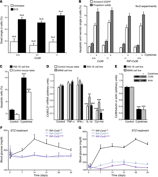
Figure 5. β cell protection requires cell contact and Cx expression.
(A) The proportion of dead cells was similar in all control islet cell suspensions. STZ similarly increased this proportion in all groups. n as indicated. (B) Islet cells of control and homozygous mice of both Cx36 and RIP-Cx36 lines showed increased apoptosis (white) and necrosis (gray) after exposure to the cytokine mix of IL-1β, IFN-γ, and TNF-α. (C) The cytokine mix increased apoptosis of INS1E cells and control C57BL/6 islets. (D) The cytokine mix decreased Cx36 mRNA in mouse islets and in MIN6 and INS-1E cells. Values are expressed relative to L27 gene level. (E) The cytokine mix also decreased Cx36 in extracts of INS1E and MIN6 cells. Values are shown relative to the tubulin signal, normalized to control. Cx36 and actin Western blot immunolabeling, from which the quantitative data were generated, is also shown (inset). **P < 0.01, ***P < 0.001 versus corresponding control. (F) STZ injection induced hyperglycemia in RIP-Cx32–/– mice, but not in RIP-Cx32+/– and RIP-Cx32+/+ littermates. (G) RIP-Cx43–/– mice also became hyperglycemic after STZ injection, whereas RIP-Cx43+/– and RIP-Cx43+/+ littermates did not. Data are mean ± SEM of 4–6 experiments (A), of 3–5 experiments (B–E), or of 3–12 mice (F and G).

