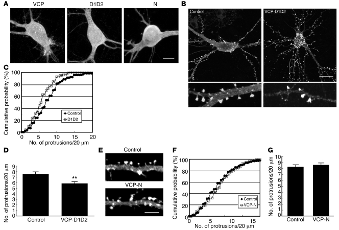
Figure 4. Overexpression of the neurofibromin-binding domain of VCP inhibits spine formation.
(A) Immunostaining using Myc-tag antibody reveals the similar expression and distribution of Myc-tagged VCP, D1D2, and N-domain in cultured hippocampal neurons. (B) Myc-tagged VCP D1D2 construct and (E) Myc-tagged N-domain and vector control were cotransfected with GFP-actin, as indicated, into cultured hippocampal neurons at 12 DIV. Six days later, the neuronal morphology was monitored by detection of GFP immunoreactivity. In B, the lower panels show enlarged images (×5.3) of the boxed regions marked in the upper panels. (C and F) Cumulative probability distributions and (D and G) graph of protrusion densities obtained from B and E, respectively. More than 22 neurons and 86 dendrites for each group of experiments were analyzed. P < 0.05, DID2 versus control; **P < 0.01. Scale bars: 10 μm (A); 20 μm (B); 5 μm (E). Values are presented as mean plus SEM.

