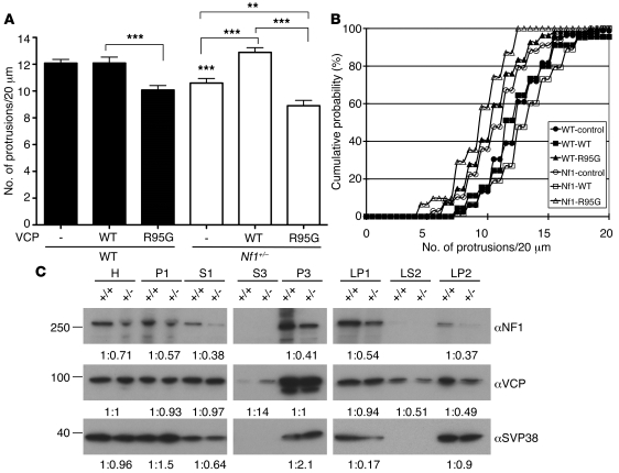
Figure 8. VCP acts downstream of neurofibromin in the regulation of dendritic spine density.
(A and B) Cultured cortical neurons isolated from Nf1+/– mice and WT littermates were cotransfected with GFP-actin and Myc-tagged WT VCP or R95G mutant or vector control at 12 DIV. The density of dendritic spines was determined at 18 DIV by staining using GFP and Myc antibodies. (A) Mean values plus SEM of protrusion density. **P < 0.01, ***P < 0.001. (B) Cumulative probability of protrusion density. In total, 19–26 neurons and 31–82 dendrites were analyzed. P < 0.01, WT R95G versus WT WT and Nf1+/– control versus WT control; P < 0.001, Nf1+/– WT versus Nf1+/– control and versus Nf1+/– R95G. (C) Subcellular distribution of VCP in Nf1+/– brain. Subcellular fractions of WT and Nf1+/– brains were isolated by a series of centrifugations (95) and analyzed by immunoblotting. Synaptophysin (SVP38) was used as a loading control of LP2. H, total homogenate; P1, crude nuclei, unbroken cells, and debris; S1, supernatant of P1; P3, light membrane fraction (including ER and Golgi body); S3, soluble cytosolic fraction; LP1, lysed synaptosomal membrane; LP2, crude synaptic vesicles; LS2, soluble synaptic cytosol. The signals were quantified using Gel-Pro Analyzer (Media Cybernetics). The intensity ratios of WT to Nf1+/– of each fraction are shown.

