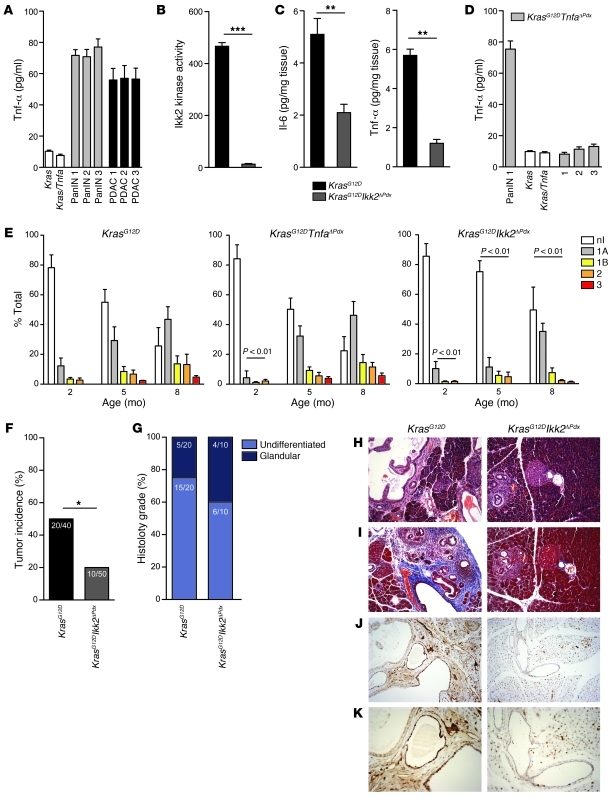
Figure 1. Genetic deletion of Ikk2 inhibits PanIN progression.
(A) Tnf-α secretion by ductal cell lines derived from KrasG12D PanIN- or PDAC-bearing mice measured by ELISA. Control cells were generated from Kras and Kras/Tnfa cre-negative pancreases. (B) Cellular Ikk2 kinase activity in cell lines derived from KrasG12D and KrasG12DIkk2ΔPdx mice. (C) Il-6 and Tnf-α secretion in pancreatic tissue of KrasG12D and KrasG12DIkk2ΔPdx mice. n = 6; **P < 0.01, ***P < 0.01. (D) Tnf-α secretion by cell lines derived from KrasG12D (PanIN 1) or KrasG12DTnfaΔPdx PanIN- or PDAC-bearing mice. Cre-negative Kras and Kras/Tnfa control cells were included. Data in C are shown as mean + SD of n = 6 mice, and data in A, B, and D are mean + SD of triplicate experiments. (E) Quantification of the proportion of pancreas occupied by PanIN lesions. Frequency and grade of the lesions was quantified at 2, 5, and 8 months of age. Data are shown as mean + SD; P < 0.01. nl, no lesion. (F) Tumor incidence and (G) histology grade in KrasG12D and KrasG12DIkk2ΔPdx mice. *P < 0.05. (H–K) KrasG12D and KrasG12DIkk2ΔPdx 4-month old pancreases stained with (H) hematoxylin and eosin, (I) Masson’s trichrome (blue, collagen; red, muscle fibers and cytoplasm; black, nuclei) and (J and K) anti-PCNA. Original magnification, ×10 (H–J), ×20 (K).

