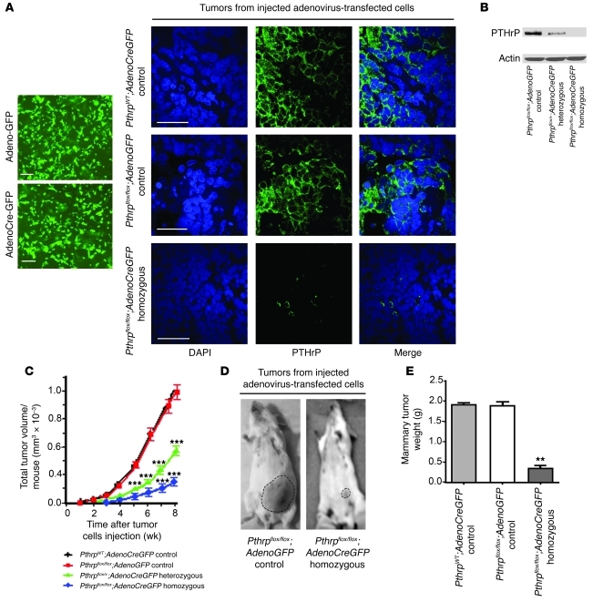
Figure 4. A more complete ablation of Pthrp by Cre-carrying adenovirus further delays breast cancer initiation and progression.
(A) Adenovirus-transfected tumor cells selected by flow cytometry: left, cell fluorescence for GFP; right, confocal images of IF staining for DAPI (blue) and PTHrP (green) in mammary tumors derived from injected adenovirus-transfected tumor cells illustrating near-complete disappearance of PTHrP expression in homozygous tumors. Scale bars: 200 μm. (B) Western blot quantifying PTHrP expression in Pthrpflox/flox tumor cells transfected with adenoGFP. Lane 1, control, Pthrpflox/flox adenoGFP; lanes 2 and 3, hetero- and homozygous, Pthrpflox/+ or Pthrpflox/flox adenoCreGFP, respectively. (C) Tumor volume per animal for tumors derived from adenovirus-transfected tumor cells injected into the MFPs of syngeneic mice. Values represent mean ± SD, n = 12 mice for each group. ***P < 0.001. (D) Tumor load in whole animals 8 weeks after adenovirus-transfected cell injection. (E) Average weight of breast tumor load per mouse at sacrifice. Values represent mean ± SD, n = 12 mice per group. **P < 0.01.

