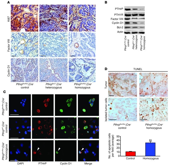
Figure 5. Pthrp ablation modifies cell-cycle, apoptosis, and angiogenesis events.
(A) IHC staining of tumor tissues at 13 weeks showing a decrease in differentiation factor Ki67 (top), angiogenesis factor VIII (middle), and cyclin D1 (bottom) with degree of Pthrp ablation. (B) Western blot illustrating no change in PTH/PTHrP receptor 1 expression, but decreases in factor VIII, cyclin D1, and Bcl-2 with degree of Pthrp ablation. (C) Confocal images of IF staining in cultured cells isolated from tumors showing colocalization of PTHrP and cyclin D1 expression. The residual cells that escaped ablation and are still capable of expressing PTHrP are the only ones expressing cyclin D1 (homozygous, bottom row, arrowheads). Shown are DAPI (blue), PTHrP (red), and cyclin D1 (green). (D) TUNEL staining of breast tumor tissue (top) or in cells isolated from tumors and cultured (bottom), showing more abundant apoptotic events in homozygous tumors. Bottom panel: histogram showing average number of apoptotic cells per field in isolated tumor cells. Scale bars: 50 μm. Values represent mean ± SD. **P < 0.01.

