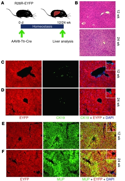
Figure 4. Hepatocyte fate tracing in liver homeostasis.
(A) Livers of adult R26R-EYFP mice were analyzed 12 and 24 weeks after injection of 4 × 10¹¹ viral genomes of AAV8-Ttr-Cre. (B) H&E staining shows normal liver histologies. (C and D) Coimmunostaining for EYFP (red) and CK19 (green) shows normal hepatocyte plates and bile ducts 12 weeks (C) and 24 weeks (D) after AAV8-Ttr-Cre injection. No hepatocyte appears EYFP negative at both time points. (E and F) Coimmunostaining for EYFP (red) and MUP (green) confirms that all hepatocytes express EYFP 12 weeks (E) and 24 weeks (F) after AAV8-Ttr-Cre injection. Nuclei were stained with DAPI (blue). Original magnification, ×100, insets ×200. 15 liver sections from 3 mice were analyzed for each experiment.

