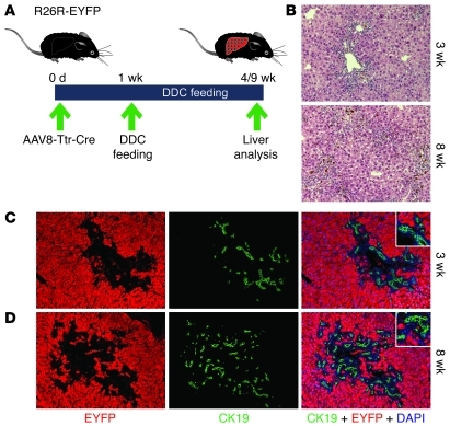
Figure 9. Hepatocyte fate tracing after DDC feeding.
(A) Adult R26R-EYFP mice were injected with 4 × 10¹¹ viral genomes of AAV8-Ttr-Cre, and DDC feeding began 1 week later. Livers were analyzed after 3 or 8 weeks of DDC feeding. (B) H&E staining shows ductular reactions expanding with time of DDC feeding. (C and D) Coimmunostaining for EYFP (red) and CK19 (green) shows no double-positive cells after 3 (C) or 8 (D) weeks of DDC feeding. Nuclei were stained with DAPI (blue). Original magnification, ×100, insets ×200. 20 liver sections from 4 mice were analyzed for each experiment.

