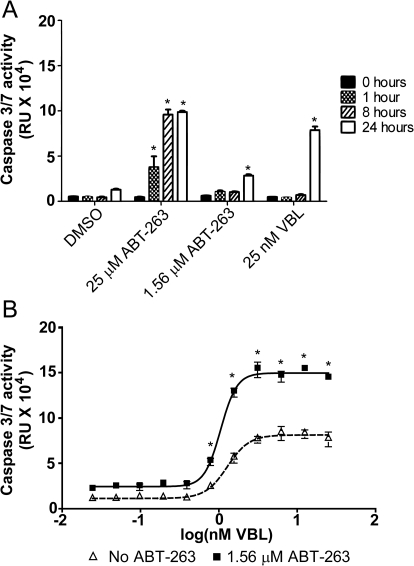
Fig. 5.
Induction of intrinsic apoptosis induced by VBL and ABT-263. A, activation of caspase-3/7 in T98G cells was measured at toxic (25 μM) and nontoxic (1.56 μM) concentrations of ABT-263 and toxic (25 nM) concentrations of VBL over time. Caspase-3/7 activation was measured at 0 (black), 1 (checkered), 8 (diagonal lines), and 24 h (Whitehurst et al., 2007). B, T98G cells were treated with increasing concentrations of VBL (25 pM to 25 nM) in the presence and absence of 1.56 μM ABT-263. Caspase-3/7 activation was measured at 24 h using a Caspase-3/7 Glo Assay kit. Δ, Cells treated with increasing concentrations of VBL. ■, Cells treated with combinations of VBL and ABT-263. Each value is the mean of three independent experiments. Bars represent S.E.M. *, p ≤ 0.05.

