Fig. 1.
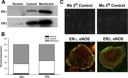
Human BECs express ERs. A, Western analyses of fractions of unstimulated BECs show that ERα and ERβ both localize to the plasma membrane and cytosol, with a small degree of expression in the nucleus. B, bar graph summarizes results from four patients. C, fluorescent immunostaining demonstrated ERα and ERβ expression in BECs, with substantial colocalization of eNOS with either isoform. An Alexa Fluor 488 dye was used to visualize ERs, whereas Cy3 was used for eNOS. Rb, rabbit; Mo, mouse. Scale bar, 10 μm (top panels), 2 μm (bottom panels).

