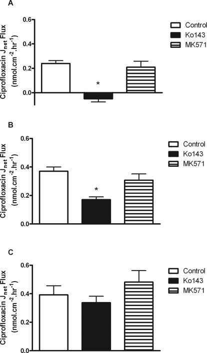
Fig. 2.
Net transepithelial [14C]ciprofloxacin flux (Jnet) across confluent monolayers of bcrp1-MDCKII cells (A), low-PA Caco-2 cells (B), and high-PA Caco-2 cells (C) grown on permeable Transwell supports. Fluxes were determined in the presence and absence of Ko143 (1 μM) and MK571 (10 μM). Other experimental details are as in the legend for Fig. 1. *, significant reductions in Jnet compared with control values, P < 0.05. n = 3 separate experiments.

