FIGURE 1:
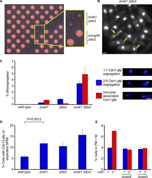
Nsk1 is required for accurate chromosome segregation and timely anaphase onset. (A) Plate containing selective antibiotics and 1 mg/ml phloxine B from a genome-wide screen for mutants that are slow growing in the absence of Dis2. Phloxin B stains dead cells. (B) Log-phase Δnsk1 Δdis2 cells were fixed and stained with DAPI and calcofluor to stain chromatin and septa, respectively. Closed arrow indicates a cell undergoing abnormal chromosome segregation in mitosis. Open arrows indicate a cell in which all chromosomes have been segregated to one of the two daughter cells. Scale bars: 2 μm. (C) G2-synchronized wild-type, Δnsk1, Δdis2, and Δnsk1 Δdis2 cells expressing lys1:lacO gfp-NLS-lacI cdc11-cfp were isolated from a 10–40% lactose gradient and segregation of chromosome 1 was monitored in the following anaphase. The percentage of missegregation was calculated as the total number of cells displaying either nondisjunction (blue bar) or non-SPB–associated sister chromatids (red bar). Error bars are the SD from the mean of three independent experiments. (D) Log-phase wild-type, Δnsk1, Δdis2, and Δnsk1 Δdis2 cells expressing Cdc13-gfp were fixed, and the percentage of cells with spindle- and SPB-associated Cdc13 assessed. Error bars are the SD from the mean of three independent experiments. (E) Log-phase cultures of wild-type, Δmad2, Δmad3, Δnsk1, Δnsk1 Δmad2, and Δnsk1 Δmad3 cells expressing dad1-gfp and sid4-tdtomato were fixed, and the percentage of prometaphase and metaphase (PM + M) cells were assessed. Error bars are the SD from the mean of three independent experiments.

