FIGURE 5:
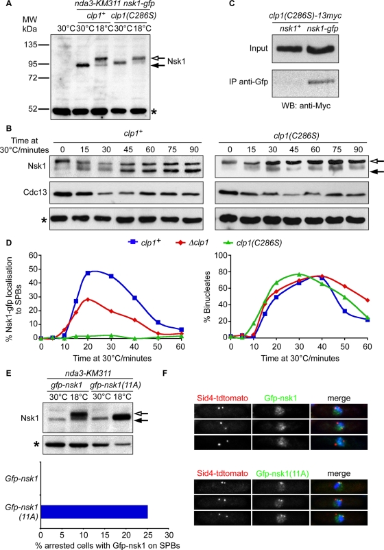
Dephosphorylation of Nsk1 by Clp1 promotes its association to the kinetochore–SPB junction. (A) Total-cell extracts were prepared from log-phase or mitotically arrested cultures of wild-type, nda3-KM311 nsk1-gfp clp1+, or nda3-KM311 nsk1-gfp clp1(C286S) cells. Proteins were separated by SDS–PAGE and Western blotting and probed with anti-GFP antibodies. Closed arrow indicates major Nsk1-gfp species observed in log-phase extracts. Open arrow indicates slower-migrating species observed in mitotically arrested extracts. Asterisk indicates a 50-kDa nonspecific band also found in the wild-type strain and used as a loading control throughout. (B) Log-phase cultures of nda3-KM311 nsk1-gfp clp1+ or nda3-KM311 nsk1-gfp clp1(C286S) cells were arrested in mitosis by incubation at 18°C for 6 h and then released to the permissive temperature. At the times indicated, total-cell extracts were prepared and probed by Western blotting using anti-GFP antibodies to detect Nsk1-gfp and anti-Cdc13 antibodies to monitor mitotic exit. Open arrow indicates slower-migrating Nsk1-gfp. Closed arrow indicates more rapidly migrating species. Some slower-migrating Nsk1-gfp remains after 90 min at permissive temperature, but note that ∼20% of cells do not enter anaphase under this regime. (C) Extracts were prepared from log-phase cultures of clp1(C286S)-13myc or nsk1-gfp clp1(C286S)-13myc cells and incubated in the presence of anti-GFP antibodies. Extracts and immunoprecipitates were probed by Western blotting using anti-Myc antibodies. (D) Log-phase cultures of nda3-KM311 nsk1-gfp sid4-tdtomato, nda3-KM311 nsk1-gfp sid4-tdtomato Δclp1, and nda3-KM311 nsk1-gfp sid4-tdtomato clp1(C286S) cells were arrested in mitosis by incubation at 18°C for 6 h and then released to the permissive temperature. At each time point, cells were fixed with formaldehyde and the percentage of cells displaying SPB-associated Nsk1 (left panel) or two separated nuclei (right panel) was measured. (E) Log-phase cultures of nmt1:nsk1-gfp Δnsk1 sid4-tdtomato nda3-KM311 or nmt1:nsk1-11A-gfp Δnsk1 sid4-tdtomato nda3-KM311 cells were grown in rich medium (containing thiamine) either at 30°C or arrested in mitosis by incubation at 18°C for 6 h. Total-cell extracts were prepared, and proteins were separated by SDS–PAGE and Western blotting and probed with anti-GFP antibodies as in (A). Alternatively, mitotically arrested cells were fixed, and the percentage of cells with Nsk1 associated to spindle poles was assessed by fluorescence microscopy. (F) Log-phase cultures of nmt1:nsk1-gfp Δnsk1 sid4-tdtomato or nmt1:nsk1-11A-gfp Δnsk1 sid4-tdtomato cells grown in rich medium (containing thiamine) at 30°C were fixed and analyzed by fluorescence microscopy.

