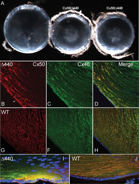
FIGURE 6:
Cx50Δ440 homozygote lenses are small, with nuclear cataracts, and display abnormal connexin distribution. (A) A comparison of Cx50Δ440 knock-in lenses (middle, right) and littermate controls (left) at P28. Knock-in lenses were smaller and developed nuclear cataracts, similar to Cx50-null mouse lines (White et al., 1998; Rong et al., 2002). (B–J) An immunofluorescence comparison of connexin distribution in P12 knock-in and littermate control lenses. (B) Cx50Δ440 was only evident in superficial cortical fibers and accumulated in large linear or irregular patches. Intracellular signal in epithelium (arrow) is higher than in control. (C) Cx46 distribution in the knock-in was abnormal in anterior epithelium and superficial fibers, but deeper fibers displayed typical plaques. (G, F, H) Cx50 and Cx46 were evenly distributed in small puncta in all fibers of the control. (I) High levels of intracellular Cx50Δ440 were evident in bow region fibers of the knock-in. (J) Intracellular connexin is not obvious in control bow region.

