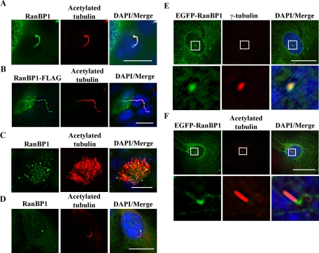
FIGURE 3:
RanBP1 localizes to the primary cilia and basal bodies. (A) RanBP1 localizes to primary cilia in IMCD3 cells. IMCD3 cells were cultured on chamberslides for 24 h in complete media and then serum starved for an additional 24 h. Cells were fixed and stained with anti–acetylated tubulin (red) and anti-RanBP1 (green). (B) RanBP1 localizes to primary cilium in MDCK cells. RanBP1-FLAG–inducible MDCK cells were grown on Transwell filters for 5 d and induced with 50 ng/ml DOX for 48 h. Cells were fixed and stained with anti-FLAG (green) and anti–acetylated tubulin (red). (C) RanBP1 localizes to motile cilia in air tract epithelia. Air tract epithelial cells were differentiated on Transwell filters for 3 wk and stained with anti-RanBP1 (green) and anti–acetylated tubulin (red). (D) RanBP1 localizes to the basal body in TERT RPE cells. RPE cells were seeded on chamber slides and cultured with serum-free media for 48 h. Cells were fixed with 4% paraformaldehyde and stained with anti-RanBP1 (green) and anti–acetylated tubulin (red). (E, F) EGFP-RanBP1 localizes to the basal body in TERT RPE cells. Inducible EGFP-RanBP1 RPE cells were seeded on chamberslides and induced with 50 ng/ml DOX for 48 h in serum-free media. Cells were fixed and stained with anti–γ-tubulin (red) in E or anti–acetylated tubulin (red) in F. Bottom, high-amplification insets from the boxed regions. Scale bars, 10 μm.

