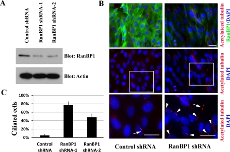
FIGURE 4:
RanBP1 knockdown initiates primary cilium formation. (A) TERT RPE cells were individually infected with two RanBP1 shRNAs and one control shRNA (luciferase shRNA), and stable pools were selected. Cell lysates were resolved by Bis-Tris PAGE and immunoblotted with the antibodies indicated. (B) TERT RPE cells stably expressing RanBP1 shRNAs or control shRNA were grown on chamberslides in complete DME/F12 growth media for 48 h. Top, samples were fixed and stained with anti–acetylated tubulin (red) and anti-RanBP1 antibodies (green). Middle, the acetylated tubulin staining individually. Bottom, high-amplification inset of the outlined region. Nuclei were visualized by DAPI staining. Arrows, cytokinetic bridges of mitotic cells; arrowheads, primary cilia. (C) Quantification of the experiments performed in B: 100 cells from each of the three cell lines were counted. The bar graph represents the mean of three individual experiments ± SD. Scale bars, 10 μm.

