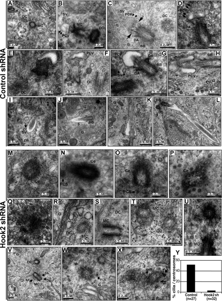
FIGURE 4:
Hook2 is involved before the formation of the pericentriolar vesicle during the initiation of ciliogenesis. (A–X) Both control shRNA 2.3 and Hook2 shRNA 2.5 clones were assayed for primary cilium formation after 7 d in culture by TEM analysis. The different steps of ciliogenesis are shown for the control cells: (A) centriole duplication, (B) pericentriolar vesicle attachment, (C, D) vesicle flattening and axoneme growth, (E–H) sheath growth, (I, J) ciliary elongation, and (K, L) ciliary maturation. Only single centrioles (M–P), as well as centriole duplication (Q–U) and maturation (V–X), were observed for the Hook2-depleted cells. BB, basal body; BFC, basal foot and cap; C, cilium; Ce, centriole; DA, distal appendages; DC, daughter centriole; E, endosome; ER, endoplasmic reticulum; MC, mother centriole; G, Golgi apparatus; N, nucleus; PCS, pericentriolar satellite; PCV, pericentriolar vesicle; S, sheath; SA, subdistal appendages; SV, secondary vesicle; μ, microtubule. Long arrows in C depict a growing axoneme that flattens a pericentriolar vesicle to form the sheath at the distal tip of the mother centriole. The shorter arrows in G and I point at a secondary vesicle (SV) fusing with the ciliary sheath. (Y) Percentage of centrosomes that were associated with a pericentriolar vesicle or a cilium in control and Hook2-depleted cells. See also Supplemental Figure S3.

