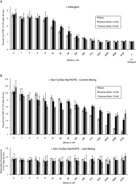
FIGURE 7:
Reactions with increasing concentrations of external biotin reveal a fusion-dependent membrane permeabilization. (A) RPLs (250 μM donors and 250 μM acceptors) were mixed in RB150 with increasing amounts of biotin (biotin [light gray columns], dextran-biotin 10,000 [black columns], or dextran-biotin 70,000 [dark gray columns]) and lysed by addition of Thesit to 0.1% (wt/vol). As control, one sample did not receive any detergent to establish the level of background signal. Shown is the relative increase of PhycoE/Cy5 FRET signal after 90 min as the average of n = 3 independent experiments ± SD values. (B) RPLs (250 μM donors and 250 μM acceptors) were mixed in RB150 with Mg2+/ATP and increasing amounts of biotin (biotin [light gray columns], dextran-biotin 10,000 [black columns], or dextran-biotin 70,000 [dark gray columns]) and incubated with Sec17p/Sec18p/HOPS at 27°C for 90 min. The relative increase of the PhycoE/Cy5 FRET signal after 90 min is displayed as the average of n = 3 independent experiments ± SD values.

