FIGURE 8:
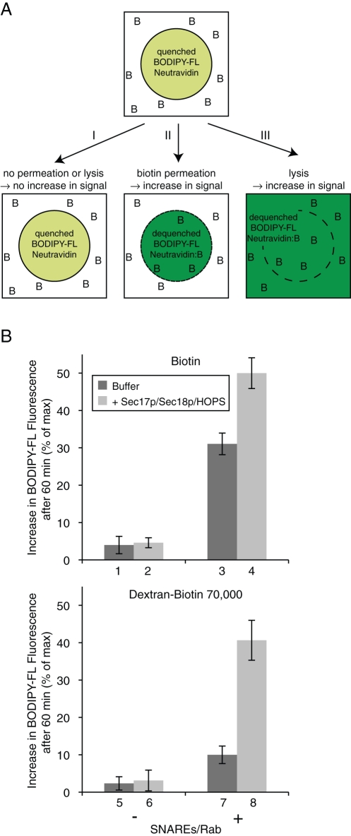
Permeation of biotin across membranes depends on the presence of SNAREs. (A) Scheme of the biotin permeation assay. Liposomes with entrapped BODIPY-FL–NeutrAvidin are incubated in the presence of biotin (as free or dextran-bound biotin; depicted as B). As long as neither permeation nor lysis occurs, the BODIPY-FL–NeutrAvidin remains in its quenched state (I). When biotin gets access to the lumenal compartment due to membrane permeation (II), or when the identity of inside and outside compartments gets lost due to lysis (III), the binding of biotin to NeutrAvidin dequenches the BODIPY-FL fluorescence signal. (B) BODIPY-FL–labeled NeutrAvidin–containing liposomes (500 μM lipid), which were either protein free (columns 1, 2, 5, and 6) or bore SNARE and Rab proteins (columns 3, 4, 7, and 8), were mixed in RB150 with Mg2+/ATP and incubated for 60 min at 27°C in the presence of biotin (top panel) or dextran-biotin 70,000 (bottom panel) with either Sec17p/Sec18p/HOPS (light gray columns) or their respective buffers (dark gray columns). The increase of BODIPY-FL fluorescence after 60 min relative to the maximal increase obtained by detergent addition is displayed as the average of n = 3 independent experiments ± SD values.

