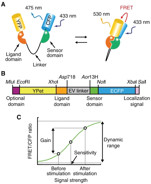
FIGURE 1:
Optimized backbone of an intramolecular FRET biosensor. (A) Mode of action of the intramolecular FRET biosensor. (B) Structure of the DNA encoding an optimized intramolecular FRET biosensor. Shown are the unique restriction enzyme sites used to exchange each domain for the development of the biosensor. (C) Schematic representation of the titration curve of FRET/CFP ratio in intramolecular FRET biosensors.

