FIGURE 6:
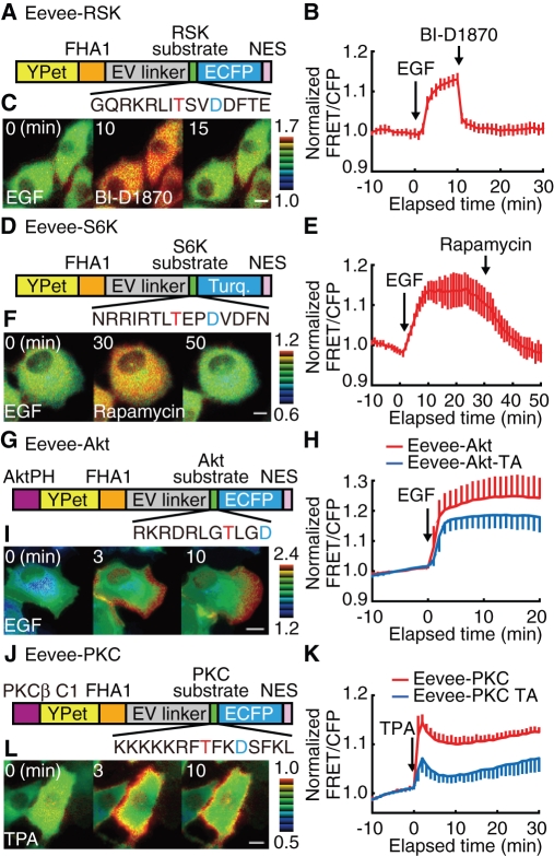
Novel Ser/Thr kinase FRET biosensors based on the Eevee backbone. (A, D, G, and J) Structures of Eevee-RSK, Eevee-S6K, Eevee-Akt, and Eevee-PKC, which are FRET biosensors of RSK, S6K, Akt, and classical PKC activities, respectively. Red and blue letters in the substrate peptide sequences denote the phosphorylation sites and amino-acid substitutions to increase the affinity to FHA1. (B, E, H, and K) HeLa cells expressing Eevee-RSK (B), Eevee-S6K (E), Eevee-PKC (K), or Cos7 cells expressing Eevee-Akt (H) were time lapse–imaged and stimulated with 10 ng/ml EGF (B and E), 50 ng/ml EGF (H), or 1 μM TPA (K). Cells expressing Eevee-RSK and Eevee-S6K were further treated with 10 nM BI-D1870 (B) and 100 nM rapamycin (E), respectively, at 30 min after EGF stimulation. The FRET/CFP ratio of each cell was normalized by dividing by the averaged FRET/CFP value before stimulation (Supplemental Videos S7–S10). The mean and SD from at least 10 cells are plotted against time. (C, F, I, and L) Representative FRET/CFP ratio images with Eevee-RSK (C), Eevee-S6K (F), Eevee-Akt (I), and Eevee-PKC (L) are shown in the intensity-modulated display mode. Scale bars are 10 μm.

