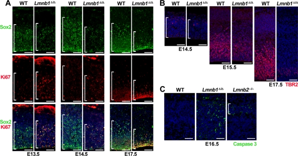
FIGURE 3:
Reduced numbers of neuronal progenitors in Lmnb1Δ/Δ brains. (A) Immunostaining for the neuronal progenitor marker Sox2 (top, green) and the mitotic antigen Ki67 (middle, red) at E13.5, E14.5, and E17.5. Bottom, merged images with DAPI (blue). Brackets mark the ventricular zone containing the Sox2+ cells. (B) Immunostaining for TBR2, a marker for intermediate neuronal progenitors, at E14.5, E15.5, and E17.5. Brackets mark the territory occupied by TBR2-positive cells (red). DNA was stained with DAPI (blue). (C) Immunostaining for active caspase 3 (green), a marker of apoptosis, in cerebral cortex of E16.5 WT, Lmnb1Δ/Δ, and Lmnb2–/– embryos. Bracket indicates the position of the cortical plate. Scale bars, A and B, 50 μm; C, 100 μm.

