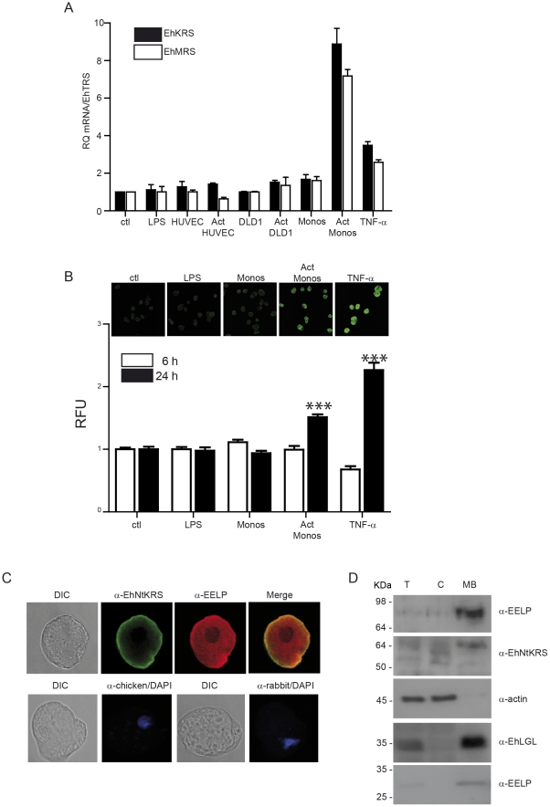
Figure 2. EhKRS and EhMRS are up-regulated by inflammation signals.
(A) qRT-PCR of EhKRS (closed bars) and EhMRS (open bars) gene expression relative to Entamoeba histolytica threonyl-tRNA synthetase (EhTRS). Trophozoites stimulated with 100 ng/ml LPS or 100 ng/ml TNF-α or co-cultured with endothelial cells (HUVEC), colonic epithelial cells (DLD1), or primary monocytes (Monos) pre-activated (Act) or not with 100 ng/ml LPS for 6 h. Values are relative to non-stimulated amoebae (ctl) and depicted as mean ± SD from at least two independent experiments performed in triplicate. (B) Quantification of EhKRS protein expression in trophozoites stimulated for 6 hours (open bars) and 24 hours (closed bars) with 100 ng/ml LPS or 100 ng/ml TNF-α or co-cultured with monocytes (Monos) or monocytes previously activated with LPS for 6 hours (Act Monos). The fluorescence units were normalized to non-stimulated (ctl) trophozoites and depicted as mean ± SD from three different experiments. The number of cells counted per each condition was n = 40. (*** p<0.0001 vs ctl). Inserts show microphotographs of immunodetection of EhKRS protein (α-NtEhKRS) in trophozoites at 24 hours of stimulation. (C) Immunolocalization of EhKRS in Entamoeba trophozoites using affinity purified antibodies against EhKRS N-terminal (α-NtEhKRS) and C-terminal (α-EELP) domains. Controls of secondary antibodies merged with DAPI are shown in bottom row. (D) EhKRS cellular localization was evaluated by immunoblot analysis of E. histolytica lysates, marked as T (whole cell lysate), C (cytoplasmic fraction), and MB (membrane bound fraction). Specific antibodies of cytoplasmic fraction (α-actin) and membrane fraction (α-LGL) were used. α-EELP antibody recognizes a full length protein (top panel) and a C-terminal cleaved product (bottom panel). α-NtEhKRS (1∶250), α-EELP (1∶50), α-LGL (1∶1000) and α-actin (1∶1000) antibody dilutions were used.

