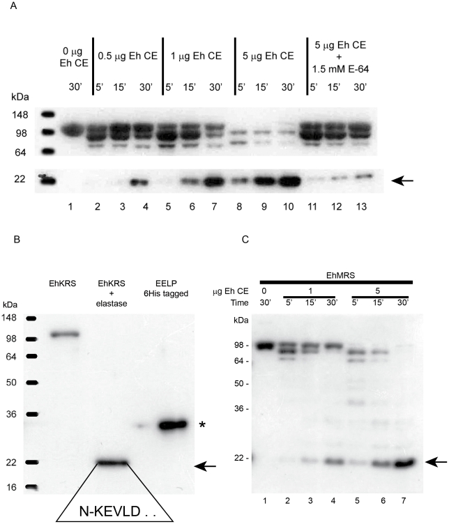
Figure 3. EhKRS protease processing.
Immunoblots analysis with α-EELP antibody of recombinant EhKRS protein digestions using Entamoeba lysates (A) or human leukocyte elastase (B). (A) 1 µg recombinant EhKRS was incubated at 37°C for 30 minutes without E. histolytica crude extract (Eh CE; lane 1) or with 0.5 µg Eh CE (lane 2–4); 1 µg Eh CE (lane 5–7); 5 µg Eh CE (lane 8–10) or 5 µg Eh CE plus protease inhibitors (lane 11–13) for 5, 15 or 30 minutes. (B) Digestion of 1 µg recombinant EhKRS protein with elastase for 30 minutes at 37°C. (C) EhMRS protease processing. Digestion of 1 µg recombinant EhMRS at 5, 15 and 30 minutes with 1 µg of Eh CE (lane 2–4) or 5 µg Eh CE (lane 5–7). Recombinant EhMRS control without Eh CE (lane 1). Digestion products were detected by immunoblot using α-EELP antibody. Arrow shows EELP product resulting from recombinant EhKRS or EhMRS. Asterisk denotes recombinant EELP with 6 His tag plus 36 amino acids at the N-terminal. Boxed sequence corresponds to the N-terminal sequence of EELP after digestion from EhKRS by elastase, as determined by Edman degradation.

