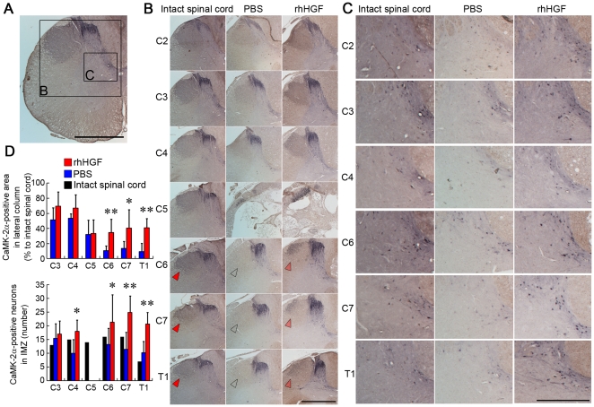
Figure 2. CaMK II α-immunoreactivity in the marmoset spinal cord.
CaMK II α-immunoreactivity (IR) in the lateral column of the white matter and in the intermediate zone (IMZ) of the gray matter in an intact spinal cord (A) and injured spinal cord 12 weeks after SCI (B, C). (A) In the intact spinal cord, CaMK II α-IR was detected in the lateral column, dorsal horn and intermediate zone of the gray matter. No CaMK II α-IR was detected in the ventral horn of the gray matter. Scale bar, 1 mm. (B) CaMK II α-IR in the lateral column, suggesting the corticospinal tract (CST) pathway, was significantly preserved even at sites caudal to the lesion epicenter (arrowheads) in the rhHGF group compared with the PBS group. Scale bar, 1 mm. (C) CaMK II α-IR was detected in cell bodies at the IMZ (laminae VI–VII), suggested to be segmental interneurons and propriospinal neurons. There were significantly more IMZ CaMK II α-positive neurons at the C4 and C6-Th1 segments in the rhHGF group than in the PBS group. IMZ CaMK2-α-positive neurons were absent from the C5 segment in both groups. Scale bar, 500 µm. (D) Quantitative analyses of the CaMK II α-IR showed significant differences between the two groups. *P<0.05, **P<0.01. (n = 6 for the rhHGF group; n = 5 for the PBS group; n = 1 for the intact spinal cord).

