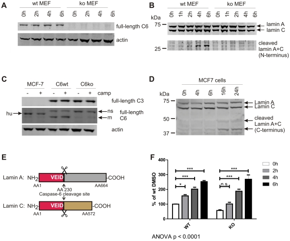
Figure 2. VEID, but not lamin A+C, is cleaved in the absence of caspase-6.
A: Caspase-6 protein (full-length, 32 kDa) is detected in MEFs generated from C6wt, but not C6ko mice. B: Endogenous lamin A protein (70 kDa) is cleaved in wt, but not C6ko MEFs after staurosporine stress for 4 h or longer. The antibody cross-reacts with full-length lamin C (60 kDa), and the cleaved band at 28 kDa has the same size for both lamin A+C (lower panel). C: MCF-7 cells express caspase-6, but not caspase-3 protein, whereas both C6wt and C6ko MEFs contain both caspases. hu: human, m: mouse, ns: non-specific band. D: MCF-7 cells were stressed with 5 µM camptothecin for different amounts of time and the cleavage of endogenous lamin A and C proteins was monitored by Western blotting with antibodies antibodies #2031 (full-length lamin A+C) and #2032 (C-terminal fragments). E: Schematic representation of lamin A and C and the caspase-6 cleavage site at AA 230. The N-terminal fragments generated by caspase-6 cleavage (red) have the same size (28 kDa) for both lamin A+C. F: C6wt or C6ko MEFs were stressed with 50 nM staurosporine for different amounts of time, lysates were generated and analyzed for cleavage of VEID-Afc. C6wt cells show a significant increase in fluorescence at each timepoint, the fluorescence signal obtained from C6ko lysates only reach a statistically significant difference from baseline after 4 h. C6ko MEFs stressed with staurosporine for 4 h or more show the same levels of VEID proteolysis as wt cells. Error bars are the SEM of N = 3 of 4 independent experiments. Statistical significance was assessed by 2-way ANOVA and post-hoc Bonferroni comparisons: *** p<0.0001, ** p<0.001, * p<0.01.

