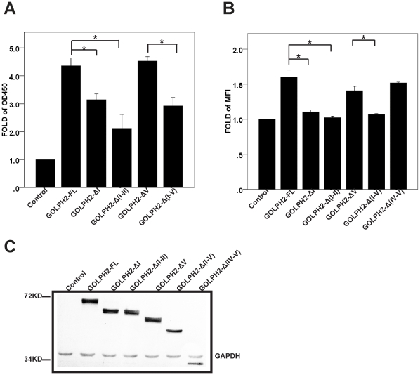
Figure 2. The TMD and cytoplasmic tail are important for GOLPH2 intracellular trafficking.
A. Secretion assay of GOLPH2 truncation proteins in HeLa cells. HeLa cells were transfected with GOLPH2 truncation expression plasmids. The medium was collected and secreted GOLPH2 was analyzed by sandwich ELISA. Data are normalized to control plasmids. Bars show mean and error bars show 95% CI of mean. n = 6, *p<0.01. B. Cell surface presentation of GOLPH2 truncation proteins in HeLa cells. About 48 h after transfection, HeLa cells were collected and resuspended. The cell surface distribution of GOLPH2 truncation proteins was analyzed by flow cytometry using anti-FLAG mAb. Data are normalized to control plasmids. Bars show mean and error bars show 95% CI of mean. n = 6, *p<0.01. C. Expression level of GOLPH2 truncation proteins. About 48 h after transfection, HeLa cells were collected and subjected to SDS PAGE followed by western blot. The expression level of GOLPH2 truncation proteins was detected with Western-blot using anti-FLAG mAb. The molecular weight marker is shown on the left.

