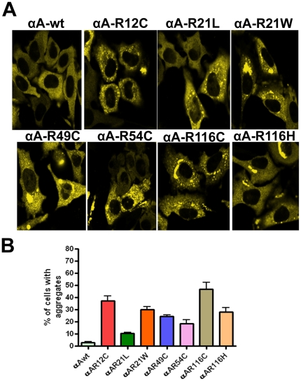
Figure 1. YFP-αA-wt and the mutant constructs, R12C, R21L, R21W, R49C, R54C, R116C and R116H were individually expressed in HeLa cells.
A: LSM Images were captured after 48 hours transfection. HeLa cells were individually transfected with 2 µg of YFP-tagged αA-wt and mutants of αA-crystallin. A homogenous expression of αA-crystallin was evident in αA-wt transfected cells. Cytoplasmic aggregates were evident in αA- crystallin mutants transfected cells. The YFP signal was excited at 514 nm and the images were collected by BP 530-600 nm filter. The images represent one of the four similar images obtained in three independent experiments. B: Graph represents per cent of cells with aggregates. The results obtained after 48 hours transfection for the individually expressed αA-wt or its mutants in HeLa cells. Cells containing aggregates were counted in 10 random fields each field with 30 cells. The mutant, R116C showed a high per cent (∼47) of cells having aggregates and the mutant R21L showed least per cent (∼10) of cells containing aggregates. The results were presented as means ± SD obtained in three independent experiments. All the mutants were statistically significant, p < 0.01.

