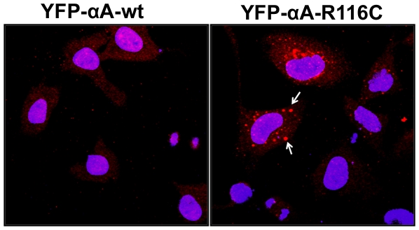
Figure 8. Overexpression of αA-crystallin mutants shows accumulation of ubiquitin inclusions in HeLa cells.
YFP-tagged αA-wt and mutant, R116C were transfected in HeLa cells. After 48 h transfection, cells were fixed and immunostained with a mouse monoclonal ubiquitin (FK2) antibody and further stained with a fluorescent conjugated secondary antibody, Alexa Flour 594 (red). The arrows indicate ubiquitinated cytoplasmic inclusions only in the mutant, R116C expressing cells. Hoechst 33342 was used to counter stain the nuclei. The images were representative of three similar images obtained in three independent experiments.

