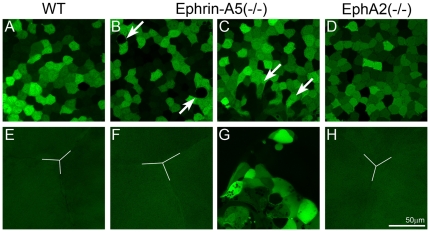
Figure 2. Confocal images of the anterior region of GFP+ wild-type (WT), ephrin-A5(-/-) and EphA2 lenses.
The WT lens shows typical mosaic GFP expression pattern in the central epithelium (A) and the Y-shape suture (Y-line) of underlying fiber cells (E). An ephrin-A5(-/-) lens displays morphological changes in a few epithelial cells (arrows in B), and the other ephrin-A5(-/-) lens shows a cluster of aberrant cells underneath the central epithelial cells (arrows in C). In addition, mislocalized aberrant cells are apparent in underlying fiber cell layers without the normal Y-shaped suture (G). The EphA2 (-/-) lens has normal central epithelial cells (D) and anterior Y-shaped suture (H). Scale bar, 50 µm.

