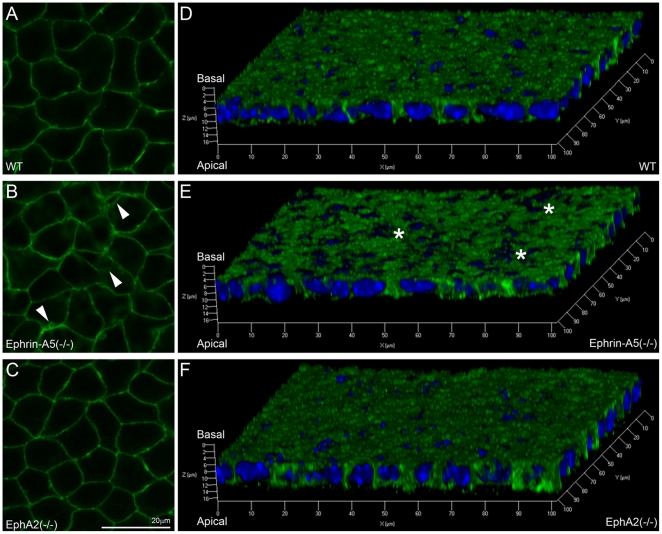
Figure 4. E-cadherin distribution in WT, ephrin-A5(-/-) and EphA2(-/-) lens capsule flat mounts.
Fluorescent images reveal normal staining signals of E-cadherin in WT and EphA2(-/-) anterior epithelial cells (A and C) but alterations in the ephrin-A5(-/-) anterior epithelium (B, indicated by arrowheads). Three-dimensional reconstructions of z-stack images labeled for E-cadherin (green) and DAPI (blue, nuclei) of lens epithelial cells from P21 WT, ephrin-A5(-/-) and EphA2(-/-) lens capsule flat mounts (D, E and F). There is a notable disruption of E-cadherin staining in ephrin-A5(-/-) anterior epithelial cells as compared to those in WT and EphA2(-/-) cells. Scale bar, 20 µm.

