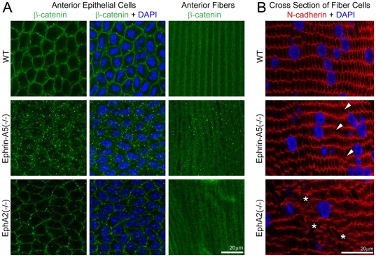
Figure 5. Beta-catenin distribution in anterior epithelial cells and underlying lens fibers and N-cadeherin localization in lens frozen sections.
Immunostaining of lens capsule flat mounts shows cell membrane distribution of β-catenin proteins in both anterior epithelial cells and fiber cells of a P21 WT lens (A). Membrane association of β-catenin remains unchanged in EphA2(-/-) anterior lens epithelial cells, but β-catenin proteins form substantial small aggregates in both anterior epithelial cells and fiber cells of ephrin-A5(-/-) lenses (A). Scale bar, 20 µm. N-cadherin is localized at the cell boundaries of hexagonal shaped lens fibers in the WT lens section (B). In the ephrin-A5(-/-) lens section, the majority of fiber cells show normal localization of N-cadherin proteins except select abnormal fiber cells (B, arrowheads). In the EphA2(-/-) lens section, the distribution of N-cadherin proteins is severely altered with some areas lacking N-cadherin staining (B, asterisks). Scale bar, 20 µm.

