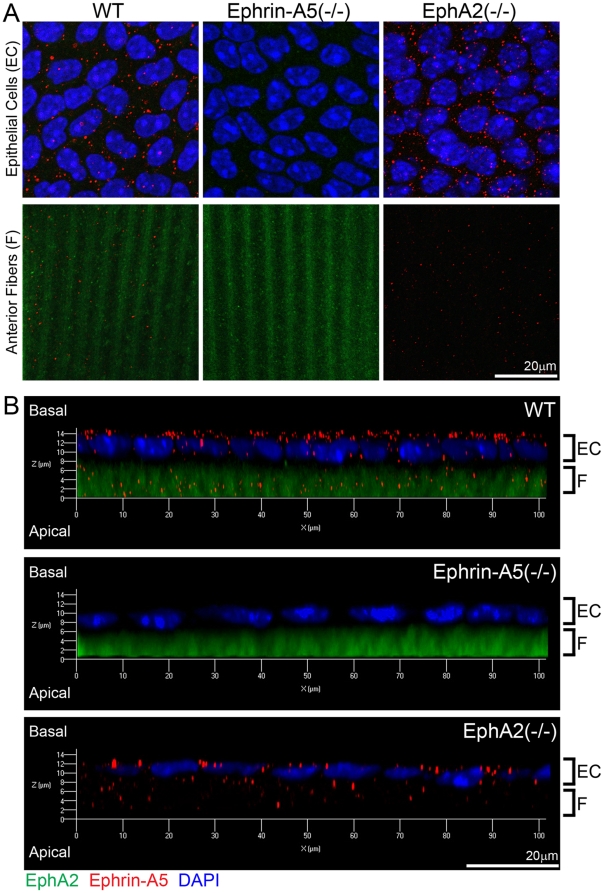
Figure 6. Localization of ephrin-A5 and EphA2 in the lens.
Double immunolabeling of ephrin-A5 (red) and EphA2 (green) with DAPI staining (blue, nuclei) of anterior lens epithelial and fiber cells from lens capsule flat mounts of P21 wild-type (WT) and ephrin-A5(-/-), EphA2(-/-) mice (A). Side views of z-stack reconstructions of anterior epithelial cells with underlying fiber cells of P21 WT, ephrin-A5(-/-), EphA2(-/-) lens capsule flat mounts reveal that ephrin-A5 proteins (red) show punctate signals mostly at the lateral and apical sides of lens epithelial cells (EC), and EphA2 proteins (green) show diffused signals in fiber cells (F) (B). Scale bar, 20 µm.

