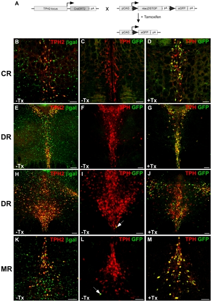
Figure 4. Inducible recombination is restricted to serotonergic neurons of adult TPH2-CreERT2/CAG-loxP.EGFP rats.
(A) TPH2-CreERT2 rats were bred to CAG-loxP.EGFP rats to generate double-transgenic TPH2-CreERT2/CAG-loxP.EGFP rats. Under uninduced baseline conditions, the loxP-flanked lacZ minigene is expressed reflecting cell-type specific CAG-promoter activity. Upon Cre-mediated recombination (+ Tamoxifen), lacZ is replaced with the second reporter gene enhanced green fluorescent protein (EGFP). The appearance of EGFP serves as an indicator of Cre mediated recombination in double transgenic rats. TPH2-CreERT2/CAG-loxP.EGFP rats were daily injected with tamoxifen (40 mg/kg) or vehicle for five consecutive days starting between P60–90. Coronal sections show dual-label fluorescence immunohistochemistry for TPH/βgal (B,E,H,K) and TPH/GFP (C,F,I,L) in vehicle-treated rats (-Tx) and TPH/GFP in tamoxifen-treated (+Tx) rats (D,G,J,M). Colocalization is visualized at the level of caudal raphe nuclei (CR) (B–D), dorsal raphe nuclei (DR) (E–J) and median raphe nuclei (MR) (K–M) using confocal images. In vehicle-treated rats, TPH2-CreERT2/CAG-loxP.EGFP rats display strong basal, non-recombined βgal expression in TPH2+ 5-HT neurons (B,E,H,K) making these rats ideally suited to monitor tamoxifen-induced Cre-mediated recombination in 5-HT neurons. (C,F,I,L) Without tamoxifen treatment, background recombination, i.e. EGFP expression (arrows) hardly occurs. (D,G,J,M) After tamoxifen treatment, the majority of TPH+ 5-HT neurons in all raphe nuclei now show EGFP expression indicating Cre-mediated recombination in these neurons (GFP+/TPH+). Scale bars: 100 µm.

