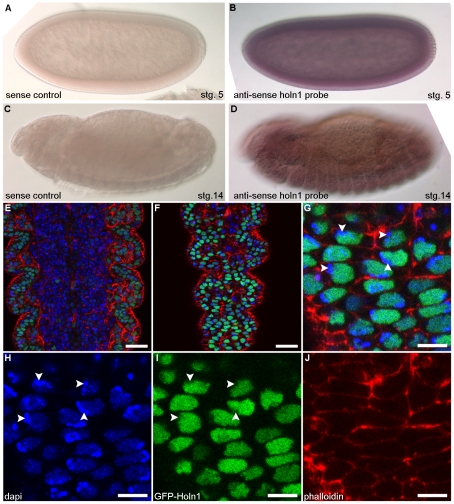
Figure 1. Expression of Holn1 in wild-type embryos.
(A–D) Expression pattern of holn1 RNA in wild-type embryos. (A,C) Sense control in situ hybridization showing lack of staining in stage 5 (A) and stage 14 (C) embryos. (B) holn1 anti-sense RNA probe shows strong maternal contribution of holn1 RNA in stage 5 embryo. (D) holn1 RNA expression is weak and ubiquitous in stage 14 embryo, enriched slightly in the nerve cord and present in the epidermis. Dorsal is to the top and anterior to the left. stg., stage. (E–J) Expression of GFP-Holn1 in the embryonic ventral epidermis under the control of the epidermal driver e22c>gal4. (E,F) GFP-Holn1 is expressed in the nuclei of ventral epidermis cells. (G–J) Magnified view of embryo shown in E,F. (E–G) Merged channels. (H) DAPI shows nuclear staining. (I) GFP-Holn1 localization in the cell nuclei. (J) Phalloidin marks filamentous actin at the cell cortex. Arrowheads in (G–I) indicate regions where heterochromatin is more condensed. All images are single Z slices. Scale bar in (E,F) = 20 µm, and in (G–J) = 10 µm.

