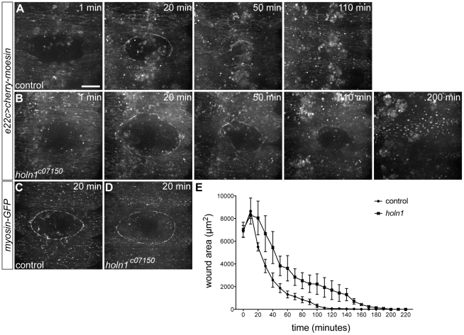
Figure 3. Time course of wound closure in control and holn1c07150 mutant embryos.
(A–B) Stills of wound closure process in e22c>cherry-moesin control (A) and e22c>cherry-moesin,holn1c07150 mutant embryos (B) at 1, 20, 50, 110, and 200 minutes after wounding. At 20 minutes after wounding, an actin cable is already formed in both control (A) and mutant embryo (B). Control embryo has its wound closed at 110 minutes after wounding, whereas mutant embryo only close around 200 minutes after wounding. (C,D) Stills of wounds in myosin-GFP control (C) and myosin-GFP,holn1c07150 mutant embryos (D) at 20 minutes after wounding. A myosin cable is observed at the wound edge in both control and mutant embryos. (E) Plot showing average wound diameter of closing wounds in control (n = 3) and holn1c07150 mutant embryos (n = 3). Both in control and mutant embryos, wounds start as 7000 µm2-diameter holes and expand in size during the first 10 minutes after wounding. After that, wound size decreases exponentially, but slower in mutants than in controls. Error bars represent the standard error of the mean for each time point. Average time of wound closure is significantly higher in mutants (194.3 minutes) compared to controls (127.7 minutes; unpaired t test, p<0.05). Images in (A–D) are projections of spinning disc confocal time-lapse recordings. Scale bar = 20 µm.

