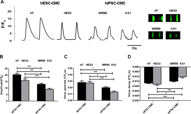
Fig. 3.
Representative tracing of calcium transients and 2-D calcium transients in a H7 & HES3 hESC derived cardiomyocytes (H7 & HES3 hESC-CMCs), IMR90 & KS1 hiPSC derived cardiomyocytes (IMR90 & KS1 hiPSC-CMCs). b Amplitude, c maximal upstroke velocity (V max upstroke), and d maximal decay velocity (V max decay) of calcium transients in the cardiomyocytes. Unpaired t-test was performed between H7 or HES3 hES-CMCs and the other two lines of iPS-CMCs (n = 10; n is corresponds to different individual cells derived from three different experiments) (*p < 0.05, **p < 0.005 and ***p < 0.0005)

